Fundus Camera
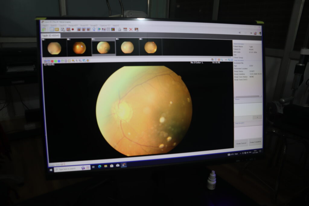
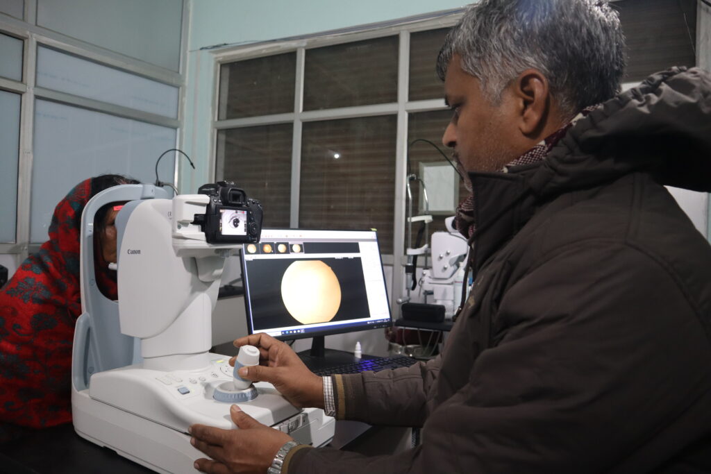
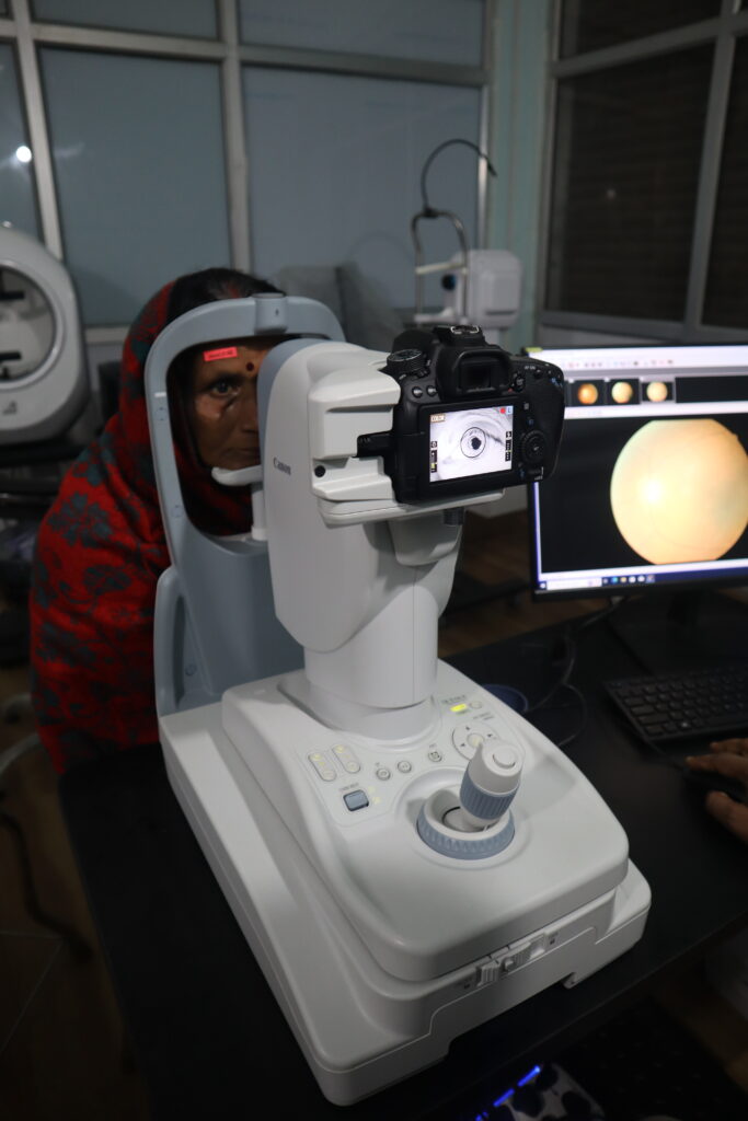
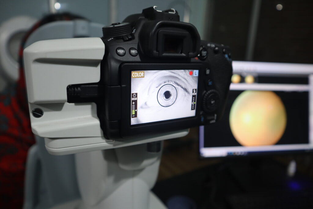
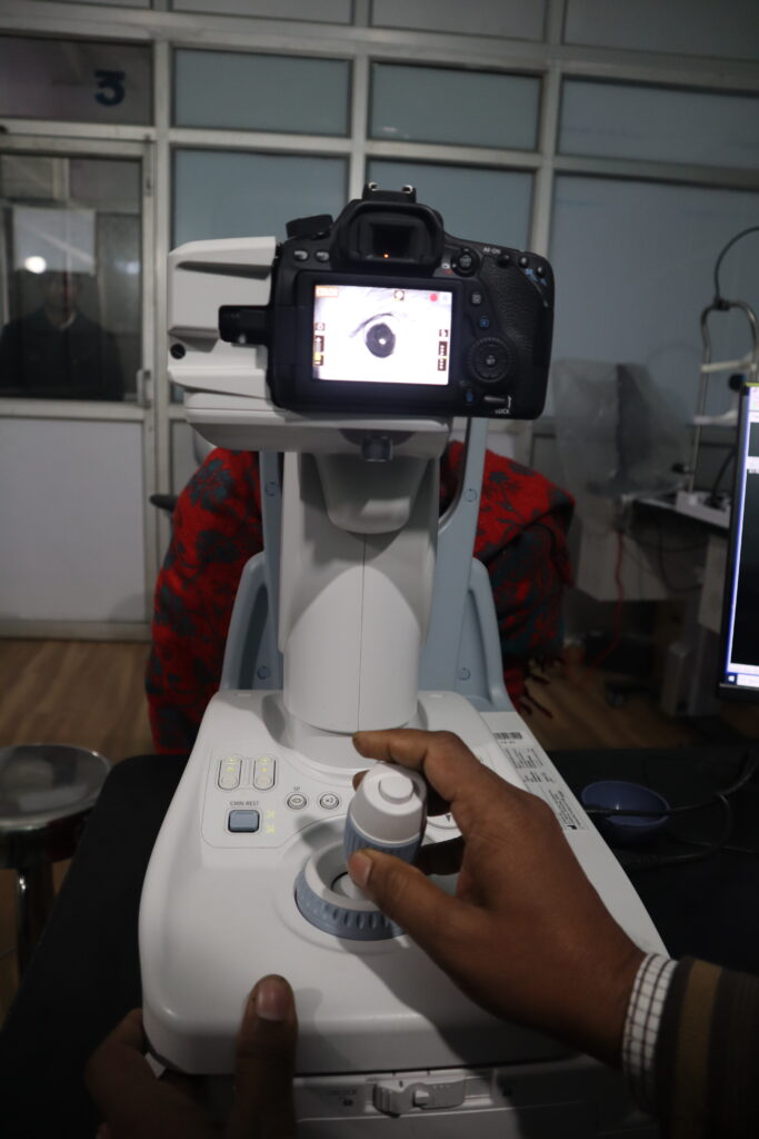
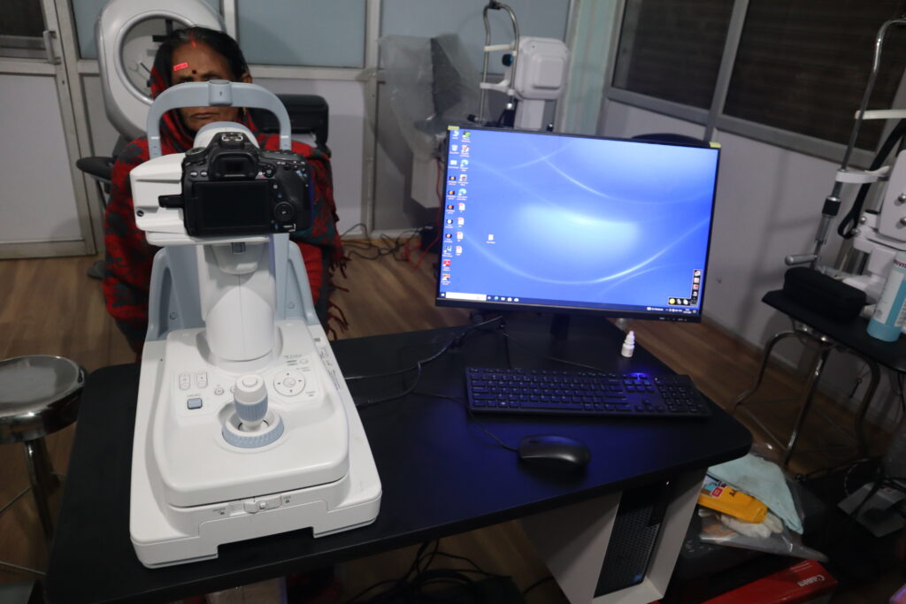
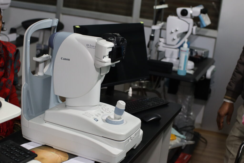
A fundus camera is a special type of camera used by eye doctors to take detailed pictures of the back part of your eye, called the retina.
Uses:
- Finding Eye Problems: It helps doctors find problems like diabetes-related eye damage, glaucoma, and macular degeneration.
- Tracking Changes: It allows doctors to see if an eye condition is getting better or worse over time.
- Recording Changes: It takes clear pictures that doctors can compare over months or years to see any changes.
- Spotting Issues Early: It can catch signs of eye issues early before they become serious.
Benefits:
- No Pain: It’s a quick and painless way to check your eyes.
- Detailed View: It gives a very detailed picture, helping doctors see tiny details they might miss with a regular exam.
- Early Detection: By finding problems early, it helps in getting timely treatment.
- Better Accuracy: It makes it easier for doctors to make accurate diagnoses.
- Comfortable for Patients: Most people find the process easy and comfortable.
फंडस कैमरा एक विशेष प्रकार का कैमरा होता है जिसका उपयोग आँखों के डॉक्टर आपकी आँखों के पीछे के हिस्से, जिसे रेटिना कहते हैं, की विस्तृत तस्वीरें लेने के लिए करते हैं।
उपयोग:
- आँखों की समस्याओं का पता लगाना: यह डॉक्टरों को मधुमेह संबंधित आँख की क्षति, ग्लूकोमा, और मैकुलर डीजनरेशन जैसी समस्याओं का पता लगाने में मदद करता है।
- परिवर्तनों को ट्रैक करना: यह डॉक्टरों को समय के साथ किसी आँख की स्थिति के बेहतर या खराब होने का पता लगाने में मदद करता है।
- परिवर्तनों को रिकॉर्ड करना: यह स्पष्ट तस्वीरें लेता है जिन्हें डॉक्टर महीनों या वर्षों बाद तुलना कर सकते हैं ताकि किसी भी परिवर्तन का पता चल सके।
- समस्याओं का प्रारंभिक चरण में पता लगाना: यह गंभीर होने से पहले आँखों की समस्याओं के संकेतों को जल्दी पकड़ सकता है।
लाभ:
- कोई दर्द नहीं: यह एक त्वरित और बिना दर्द के आँखों की जाँच करने का तरीका है।
- विस्तृत दृश्य: यह बहुत विस्तृत तस्वीरें देता है, जिससे डॉक्टर उन छोटे विवरणों को देख सकते हैं जो सामान्य जांच में छूट सकते हैं।
- प्रारंभिक पहचान: समस्याओं का प्रारंभिक चरण में पता लगाकर, यह समय पर उपचार में मदद करता है।
- बेहतर सटीकता: यह डॉक्टरों को सही निदान करने में मदद करता है।
- मरीजों के लिए आरामदायक: अधिकांश लोग इस प्रक्रिया को सरल और आरामदायक पाते हैं।
संक्षेप में, एक फंडस कैमरा डॉक्टरों को आपकी आँख के अंदर की साफ तस्वीरें लेने में मदद करता है ताकि आपकी दृष्टि स्वस्थ बनी रहे।
Auto perimeter
What Is Field Vision?
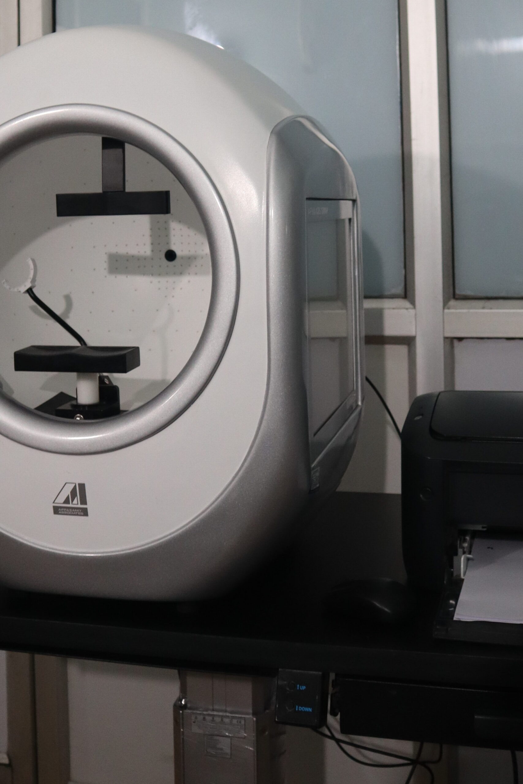
Field vision, also known as the visual field, refers to the entire area that you can see when your eyes are fixed in one position, looking straight ahead. This includes not only what you see directly in front of you but also what you see on the sides (peripheral vision).
Why Field Vision is Important:
- Comprehensive View: It gives a complete picture of what you can see without moving your eyes or head.
- Detection of Abnormalities: Helps in detecting blind spots or areas where your vision is limited, which can be early signs of eye conditions.
- Daily Activities: Essential for many daily activities like driving, reading, and navigating spaces safely.
- Health Monitoring: Changes in your visual field can indicate various health issues, not just related to the eyes but also neurological conditions.
Understanding your field vision is crucial for maintaining overall eye health and ensuring that any potential issues are detected and treated early.
दृष्टि क्षेत्र, जिसे विजुअल फील्ड भी कहा जाता है, से तात्पर्य उस पूरे क्षेत्र से है जिसे आप अपनी आँखें एक स्थिति में रखते हुए देख सकते हैं, सीधे आगे देखते हुए। इसमें न केवल आप जो सीधे सामने देखते हैं, बल्कि जो आप पक्षों पर (परिधीय दृष्टि) देखते हैं, वह भी शामिल है।
दृष्टि क्षेत्र क्यों महत्वपूर्ण है:
- व्यापक दृश्य: यह बिना अपनी आँखें या सिर घुमाए आप जो देख सकते हैं, उसका पूरा चित्र देता है।
- विकृतियों का पता लगाना: यह अंधे धब्बे या ऐसे क्षेत्रों का पता लगाने में मदद करता है जहाँ आपकी दृष्टि सीमित है, जो नेत्र स्थितियों के प्रारंभिक संकेत हो सकते हैं।
- दैनिक गतिविधियाँ: ड्राइविंग, पढ़ने और सुरक्षित रूप से स्थानों में नेविगेट करने जैसी कई दैनिक गतिविधियों के लिए आवश्यक है।
- स्वास्थ्य की निगरानी: आपके दृष्टि क्षेत्र में परिवर्तन विभिन्न स्वास्थ्य समस्याओं का संकेत दे सकते हैं, केवल आँखों से संबंधित नहीं, बल्कि न्यूरोलॉजिकल स्थितियों से भी।
आपके दृष्टि क्षेत्र को समझना समग्र नेत्र स्वास्थ्य को बनाए रखने और किसी भी संभावित समस्याओं का प्रारं
An auto perimeter is a device used by eye doctors to test your field of vision. Here’s how it helps and its benefits in simple terms:
Uses:
- Testing Vision Field: It measures how well you can see in all directions around you, not just straight ahead.
- Detecting Eye Issues: Helps identify problems like glaucoma or damage to the optic nerve by checking for blind spots and other vision abnormalities.
- Monitoring Eye Health: Tracks any changes in your vision over time to see if your condition is getting better or worse.
- Guiding Treatment: Provides detailed information that helps doctors decide the best treatment plan for you.
Benefits:
- Quick and Easy: The test is fast and simple, usually done in a few minutes.
- Accurate Results: Gives precise measurements of your vision field, helping doctors make accurate diagnoses.
- Early Detection: Helps catch eye problems early, which can lead to better treatment outcomes and prevent serious vision loss.
- Patient Comfort: The test is generally comfortable and non-invasive, meaning it doesn’t involve any pain or discomfort.
In short, an auto perimeter helps doctors get a complete picture of your vision, ensuring that any problems are detected early and treated effectively.
बिल्कुल! यहाँ हिंदी में अनुवाद:
ऑटो परिमीटर एक उपकरण है जिसका उपयोग नेत्र चिकित्सक आपके दृष्टि क्षेत्र का परीक्षण करने के लिए करते हैं। यह कैसे मदद करता है और इसके लाभ नीचे सरल शब्दों में दिए गए हैं:
उपयोग:
- दृष्टि क्षेत्र का परीक्षण: यह मापता है कि आप अपने चारों दिशाओं में कितना अच्छा देख सकते हैं, न कि केवल सीधे आगे।
- आँखों की समस्याओं का पता लगाना: यह ग्लूकोमा या ऑप्टिक नस के नुकसान जैसी समस्याओं की पहचान करने में मदद करता है, जैसे कि ब्लाइंड स्पॉट और अन्य दृष्टि असामान्यताएं।
- नेत्र स्वास्थ्य की निगरानी: यह समय के साथ आपकी दृष्टि में किसी भी परिवर्तन को ट्रैक करता है ताकि यह देखा जा सके कि आपकी स्थिति बेहतर हो रही है या नहीं।
- उपचार का मार्गदर्शन: यह विस्तृत जानकारी प्रदान करता है जो डॉक्टरों को आपके लिए सबसे अच्छा उपचार योजना निर्धारित करने में मदद करता है।
लाभ:
- तेज और आसान: यह परीक्षण तेजी से और सरलता से होता है, आमतौर पर कुछ ही मिनटों में।
- सटीक परिणाम: यह आपके दृष्टि क्षेत्र के सटीक माप देता है, जिससे डॉक्टर सटीक निदान कर सकते हैं।
- प्रारंभिक पहचान: यह आँखों की समस्याओं का जल्द पता लगाने में मदद करता है, जिससे बेहतर उपचार परिणाम मिल सकते हैं और गंभीर दृष्टि हानि को रोका जा सकता है।
- रोगी आराम: यह परीक्षण आम तौर पर आरामदायक और बिना किसी दर्द या असुविधा के होता है।
उम्मीद है यह आपकी मदद करेगा! अगर आपके पास और कोई सवाल हैं, तो बेझिझक पूछिए!
Green Lazer
Uses:
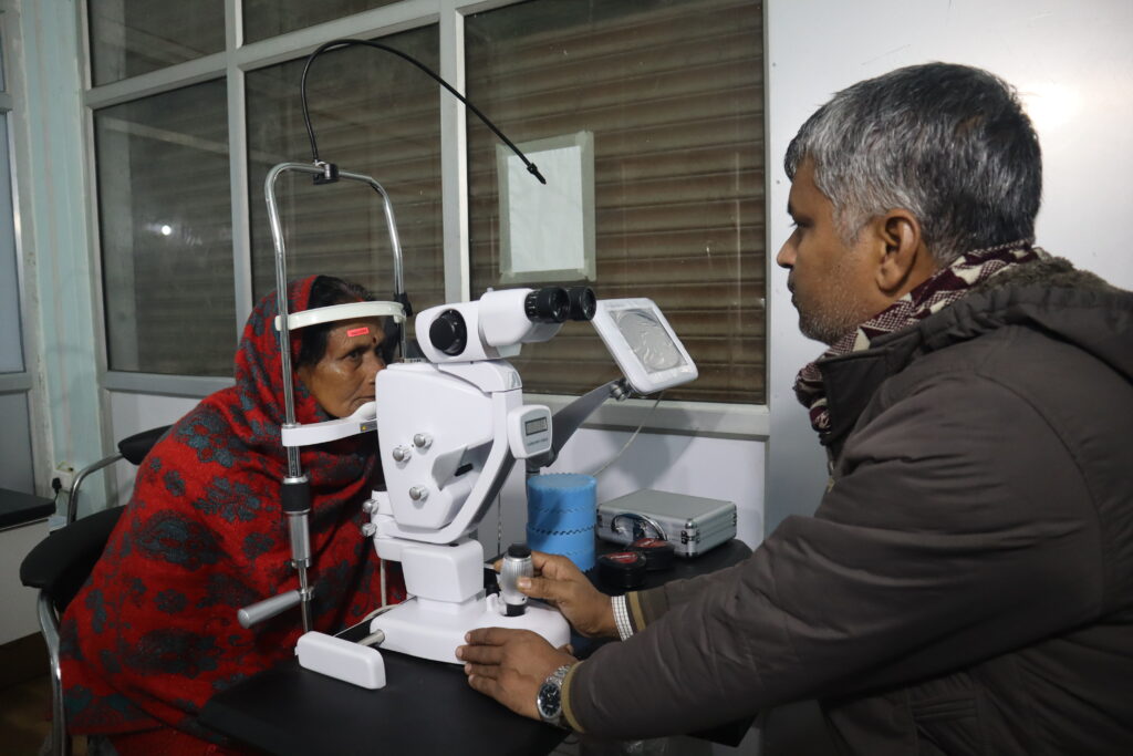
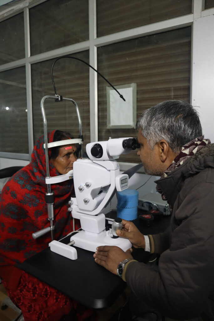
- Treating Diabetic Retinopathy: Helps in treating abnormal blood vessels caused by diabetes in the retina.
- Managing Macular Edema: Reduces swelling in the central part of the retina, improving vision.
- Sealing Retinal Tears: Seals small tears or holes in the retina to prevent further damage.
- Reducing Ischemia: Treats areas of poor blood flow in the retina to prevent further complications.
Benefits:
- Improved Vision: Many patients experience better vision after treatment.
- Non-Invasive: The procedure is done without surgery, making it less risky and more comfortable.
- Quick Recovery: Patients usually recover quickly with minimal discomfort.
- Prevents Further Damage: Helps in preventing further deterioration of the retina and potential vision loss.
यहाँ हरे लेजर के रेटिना उपचार के उपयोग और इसके लाभों का सरल शब्दों में हिंदी में अनुवाद:
उपयोग:
- डायबिटिक रेटिनोपैथी का इलाज: यह मधुमेह के कारण रेटिना में असामान्य रक्त वाहिकाओं का इलाज करने में मदद करता है।
- मैकुलर एडिमा का प्रबंधन: यह रेटिना के केंद्रीय हिस्से में सूजन को कम करता है, जिससे दृष्टि में सुधार होता है।
- रेटिनल आँसू को सील करना: यह रेटिना में छोटे आँसू या छेद को सील करता है ताकि आगे की क्षति को रोका जा सके।
- रक्त प्रवाह में कमी का इलाज: यह रेटिना में खराब रक्त प्रवाह वाले क्षेत्रों का इलाज करता है ताकि आगे की जटिलताओं को रोका जा सके।
लाभ:
- दृष्टि में सुधार: कई मरीज इलाज के बाद बेहतर दृष्टि का अनुभव करते हैं।
- गैर-आक्रामक: यह प्रक्रिया बिना सर्जरी के होती है, जिससे यह कम जोखिमपूर्ण और अधिक आरामदायक होती है।
- त्वरित रिकवरी: मरीज आमतौर पर तेजी से और कम असुविधा के साथ ठीक हो जाते हैं।
- आगे की क्षति को रोकता है: यह रेटिना के और अधिक खराब होने और संभावित दृष्टि हानि को रोकने में मदद करता है।
उम्मीद है यह आपकी मदद करेगा! अगर आपके पास और कोई सवाल हैं, तो बेझिझक पूछिए!
B-scan Ultrasound?
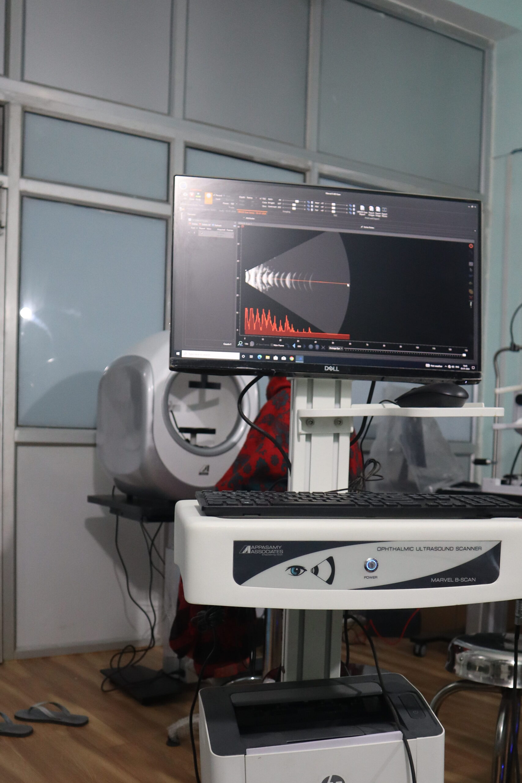
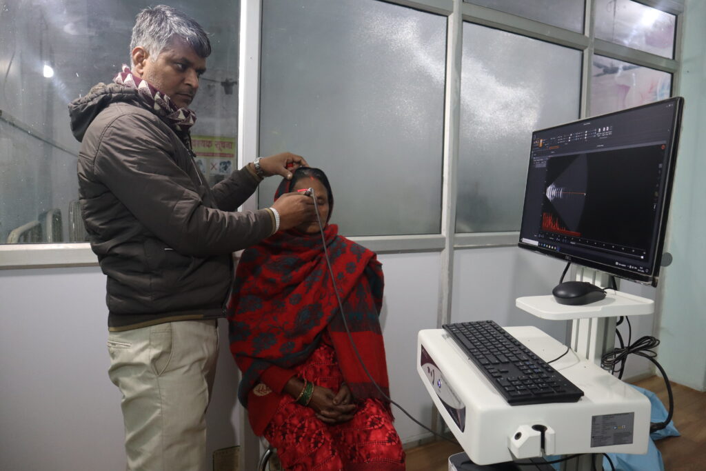
What is a B-scan Ultrasound?
A B-scan ultrasound is a non-invasive imaging test that uses high-frequency sound waves to create detailed images of the inside of your eye. It helps doctors see structures like the retina, optic nerve, and vitreous body, even if they can’t be seen directly due to opacities (like cataracts).
Uses:
- Diagnosing Eye Conditions: Helps detect issues like retinal detachment, vitreous hemorrhage, and tumors.
- Pre-Surgery Evaluation: Used before eye surgeries, especially when the view of the eye’s internal structures is blocked.
- Monitoring Eye Health: Tracks changes in the eye over time to monitor conditions like glaucoma or diabetic retinopathy.
- Guiding Treatment: Provides detailed images that help doctors plan and execute treatments more effectively.
Benefits:
- Non-Invasive: The test is safe and painless, without the need for needles or incisions.
- Detailed Visualization: Produces clear images of the eye’s internal structures, aiding in accurate diagnosis.
- Quick and Comfortable: The procedure is quick, usually taking just a few minutes, and is comfortable for patients.
- Dynamic Evaluation: Allows real-time viewing of the eye, helping doctors assess conditions more effectively.
बिल्कुल! यहाँ हिंदी में अनुवाद:
बी-स्कैन अल्ट्रासाउंड क्या है?
बी-स्कैन अल्ट्रासाउंड एक गैर-आक्रामक इमेजिंग परीक्षण है जो उच्च-आवृत्ति ध्वनि तरंगों का उपयोग करके आपकी आँख के अंदर की विस्तृत तस्वीरें बनाता है। यह डॉक्टरों को रेटिना, ऑप्टिक नस, और विट्रियस बॉडी जैसी संरचनाओं को देखने में मदद करता है, भले ही उन्हें सीधे नहीं देखा जा सकता हो (जैसे मोतियाबिंद के कारण)।
उपयोग:
- आँखों की स्थिति का निदान: रेटिनल डिटेचमेंट, विट्रियस हेमरेज, और ट्यूमर्स जैसी समस्याओं का पता लगाने में मदद करता है।
- प्र-सर्जरी मूल्यांकन: नेत्र सर्जरी से पहले उपयोग किया जाता है, विशेष रूप से जब आँख की आंतरिक संरचनाओं का दृश्य अवरुद्ध होता है।
- आँखों के स्वास्थ्य की निगरानी: समय के साथ आँखों में होने वाले परिवर्तनों को ट्रैक करता है ताकि ग्लूकोमा या डायबिटिक रेटिनोपैथी जैसी स्थितियों की निगरानी की जा सके।
- उपचार का मार्गदर्शन: विस्तृत चित्र प्रदान करता है जो डॉक्टरों को अधिक प्रभावी ढंग से उपचार की योजना बनाने और उसे निष्पादित करने में मदद करता है।
लाभ:
- गैर-आक्रामक: यह परीक्षण सुरक्षित और दर्द रहित है, जिसमें सुइयों या चीरे की आवश्यकता नहीं होती।
- विस्तृत दृश्य: आँख की आंतरिक संरचनाओं के स्पष्ट चित्र बनाता है, जो सटीक निदान में सहायता करता है।
- तेज और आरामदायक: प्रक्रिया तेजी से होती है, आमतौर पर कुछ ही मिनटों में, और मरीजों के लिए आरामदायक होती है।
- गतिशील मूल्यांकन: आँख का वास्तविक समय में अवलोकन करने की अनुमति देता है, जिससे डॉक्टर अधिक प्रभावी ढंग से स्थितियों का मूल्यांकन कर सकते हैं।
उम्मीद है यह आपकी मदद करेगा! अगर आपके पास और कोई सवाल हैं, तो बेझिझक पूछिए!
Get The Best Care For Your Eyes :
” Eyes Are Precious “
Primary Contact Details:
- Ayodhya Phaco Center
- Manager
- Ramji Gupta
- Contact No : 7235022000
- [email protected]
- Sant Kabir Eye Hospital
- Manager
- Akhand Singh
- Contact No : 8604152909
- [email protected]
- Sunbeam Eye Hospital
- Manager
- Afasar Mirza
- Contact No : 9670500097
- [email protected]
